4,651 results

- Digital Images
- Online
Illustration of the DNA double helix. The sugar-phosphate backbone of the two complementary strands are visible (red and blue).
Susan Lockhart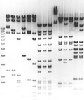
- Digital Images
- Online
Separation of DNA fragments by electrophoresis through an agarose gel. An electric current is passed through the gel and separates the DNA fragments according to size. The mixture of fragments is applied to a well at the top of the gel before the current is started. The smaller fragments travel further and reach the bottom of the gel while the larger ones remain towards the origin.
Guy Tear
- Digital Images
- Online
Cynoglossum officinale L. Boraginaceae. Houndstongue. Distribution: Europe. Culpeper (1650) writes: “... being roasted and laid to the fundament, helps the haemorrhoids. It is also good against burnings and scaldings.” It contains hepatocarcinogenic pyrrolizidine alkaloids and while people are known to eat the young leaves as a vegetable, this is inadvisable. The whole plant is hairy and may cause contact dermatitis. The use of herbal remedies, which contain these alkaloids, by the Bantu of southern Africa correlates with their high incidence of tumours of the liver and pancreas. Photographed in the Medicinal Garden of the Royal College of Physicians, London.
Dr Henry Oakeley
- Digital Images
- Online
Trichuris muris is a parasitic nematode affecting mice. Following ingestion, T. muris eggs hatch in the large intestine where they develop into adults. The anterior end of the worm burrows into the lining of the gut, leaving the posterior end protruding into the lumen of the gut. The worms mate in this orientation, and the resulting eggs are released in to the gut lumen and shed faecally.
David Goulding, Wellcome Trust Sanger Institute
- Digital Images
- Online
HeLa cell in prometaphase. The chromatin is stained red and the microtubules forming the spindle stained green. The chromatin has condensed but there is not yet complete attachment of the chromosomes to the spindle.
Matthew Daniels
- Digital Images
- Online
Human cell in interphase showing the tubulin component of the cytoskeleton in green and the DNA in red. The centrosome, to which the microtubules attach, can be seen to the right of the nucleus.
Matthew Daniels
- Digital Images
- Online
The Thorn Spider (Gasteracantha cancriformis) is a neotropical spider of the Micrathena schreibersi species of orb weavers (Araneidae). The spider has a wide distribution throughout Central and South America. Females are large and brightly colored, and have a triangular abdomen with black margins and 10 prominent spines. Males are smaller and less conspicuous than females and are less frequently encountered.
Macroscopic Solutions
- Digital Images
- Online
Fuchsia magellanica Lam. Onagraceae. Hardy fuchsia. Semi-hardy shrub. Distribution: Mountainous regions of Chile and Argentina where they are called 'Chilco' by the indigenous people, the Mapuche. The genus was discovered by Charles Plumier in Hispaniola in 1696/7, and named by him for Leonhart Fuchs (1501-1566), German Professor of Medicine, whose illustrated herbal, De Historia Stirpium (1542) attempted the identification of the plants in the Classical herbals. It also contained the first accounts of maize, Zea mays, and chilli peppers, Capsicum annuum, then recently introduced from Latin America. He was also the first person to publish an account and woodcuts of foxgloves, Digitalis purpurea and D. lutea. The book contains 500 descriptions and woodcuts of medicinal plants, arranged in alphabetical order, and relied heavily on the De Materia Medica (c. AD 70) of Dioscorides. He was a powerful influence on the herbals of Dodoens, and thence to Gerard, L’Escluse and Henry Lyte. A small quarto edition appeared in 1551, and a two volume facsimile of the 1542 edition with commentary and selected translations from the Latin was published by Stanford Press in 1999. The original woodcuts were passed from printer to printer and continued in use for 232 years (Schinz, 1774). Photographed in the Medicinal Garden of the Royal College of Physicians, London.
Dr Henry Oakeley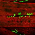
- Digital Images
- Online
Neuromuscular junction showing the association of the nerves and the muscle fibres. This image is taken from a Drosophila larva and shows the neuromuscular junctions stained with green fluorescence protein (GFP) in association with the muscle fibres in red. The muscle is stained with an antibody which provides the red colour and also shows the striated pattern of the contractile filaments (sarcomers) of the muscle.
Hermann Aberle, University of Munster
- Digital Images
- Online
Human chromosomes in early anaphase. At this stage the chromosomes have started to separate from each other and move towards opposite poles of the cell. The chromatin appears grey and the kinetochores are pink.
Matthew Daniels
- Digital Images
- Online
Human chromosomes in telophase. The chromosomes have separated and decondensed, and the new nuclear envelope forms.
Matthew Daniels
- Digital Images
- Online
Neurons or nerve cells are the core components of the brain and spinal cord of the central nervous system (CNS), and of the ganglia of the peripheral nervous system (PNS) controlling many motor and sensory functions of the body.
Odra Noel
- Digital Images
- Online
Neurons or nerve cells are the core components of the brain and spinal cord of the central nervous system (CNS), and of the ganglia of the peripheral nervous system (PNS) controlling many motor and sensory functions of the body.
Odra Noel
- Digital Images
- Online
March of the VEGF-B(10-108)-VEGFR-1D2 molecules
K R Acharya
- Digital Images
- Online
The impact of biotechnology on the world
Dan Salaman
- Digital Images
- Online
Arctium lappa L. Asteraceae. Greater Burdock. Distribution: Europe to India and Japan. Dioscorides (Beck, 2003) writes: '... helps those who spit blood and who suffer from abscesses ... plastered on it stems the pains around the joints that stem from twistings. The Leaves are applied beneficially on old ulcers.' Culpeper (1650) writes: ‘Burdanae, etc. Of Bur, Clot-Bur or Burdock, ... helps such as spit blood and matter, bruised and mixed salt and applied to the place, helpeth the bitings of mad dogs. It expels wind, easeth pains of the teeth, strengthens the back, helps the running of the reins, and the whites in women, being taken inwardly.’ The roots contain inulin, which is made into a non-digestible sweetener for diabetics. It has a multitude of uses in herbal medicine, in particular it is a component of a compound called ‘essiac’ that has been widely used as a treatment of cancers in the USA, but which is of no proven benefit. The young roots can be eaten raw or cooked. The seeds are hairy and care should be taken when harvesting them as inhaled they are reported as ‘toxic’. The root is licensed for use in Traditional Herbal Medicines in the UK (UK Medicines and Healthcare Products Regulatory Agency (MHRA)). Photographed in the Medicinal Garden of the Royal College of Physicians, London.
Dr Henry Oakeley
- Digital Images
- Online
Rudbeckia triloba L. Asteraceae Orange Cone flower. Herbaceous perennial. Distribution: North America. It is named for Olof Rudbeck, father (1630–1702) and son (1660–1740). Olof Rudbeck the Elder was professor of medicine at Uppsala University, and established a botanic garden there. He was the discoverer of the human lymphatic system. His son succeeded his father as professor of medicine, and one of his students was Carl Linnaeus (1707–88) who named the genus Rudbeckia after him and his father. It is a plant which is poisonous to cattle, sheep and pigs with no medicinal uses. Austin (1974) discusses R. hirta, also regarded as a toxic plant. It was used externally by the Cherokee to bathe sores and snakebites and made into a tea for treating diarrhoea. The Seminoles used it for headaches and fever and the Miccosukee for sunstroke and headache. The Cherokee and the Iroquois used it to treat intestinal worms Photographed in the Medicinal Garden of the Royal College of Physicians, London.
Dr Henry Oakeley
- Digital Images
- Online
Iris unguicularis Poir. Iridaceae. Algerian iris. Rhizomatous perennial. Distribution: NW Africa, E. Mediterranean It has scientifically-based potential. The rhizomes contain the chemical kaempferol which inhibits the enzyme alpha-glucosidase in the gut, reducing the rate of glucose absorption. This could be used to prevent the dangerous peaks of blood sugar that occur in diabetics and reduce eye and kidney complications. The unprocessed rhizome contains iridin, a toxic glycoside, which causes 'nausea, vomiting, diarrhoea and skin irritation'. Photographed in the Medicinal Garden of the Royal College of Physicians, London.
Dr Henry Oakeley
- Digital Images
- Online
Normal spleen showing B cells and T cells
Peter Lane and Fiona McConnell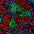
- Digital Images
- Online
Normal spleen showing B cells and T cells
Peter Lane and Fiona McConnell
- Digital Images
- Online
Normal spleen showing B cells and T cells
Peter Lane and Fiona McConnell
- Digital Images
- Online
Normal spleen showing B cells and T cells
Peter Lane and Fiona McConnell